Occlusal Pattern Assessment: Methods and Treatment Approaches
Introduction
The assessment of the occlusal pattern is a detailed clinical process that involves observing the relationship between the upper and lower teeth, both at rest and during mandibular movements. The main methods and steps include:
Clinical Examination
- The dentist observes the alignment of the teeth and the occlusal fit in a centralized bite position (centric occlusion).
- Dental contacts are checked in different mandibular movements, such as lateralization, protrusion (forward movement), and retrusion (backward movement).
- The presence of dental wear, fractures, and other alterations may indicate occlusal imbalances.
Articulation Test
- The use of articulating paper is common to verify contact points between the teeth. When biting on this paper, marks are left indicating the areas of contact. These marks help identify premature contacts or areas of excessive force.
Reference Position Examination
- The reference position refers to the most posterior position of the mandible in relation to the skull, without dental contact and without joint compression.
- This position should be comfortable for a patient with an ideal occlusion and is where stable contact occurs between the teeth and the TMJ (temporomandibular joint). The dentist evaluates whether the reference position coincides with habitual occlusion (normal bite) or if there is any deviation.
Functional Occlusion Assessment
- Functional occlusion is observed during chewing and speech movements. The dentist checks for dental interferences that may hinder the harmonious functioning of the mandible.
- Occlusal interferences occur when a tooth contacts earlier than others during mandibular movements, potentially causing wear, muscle pain, or TMJ overload.
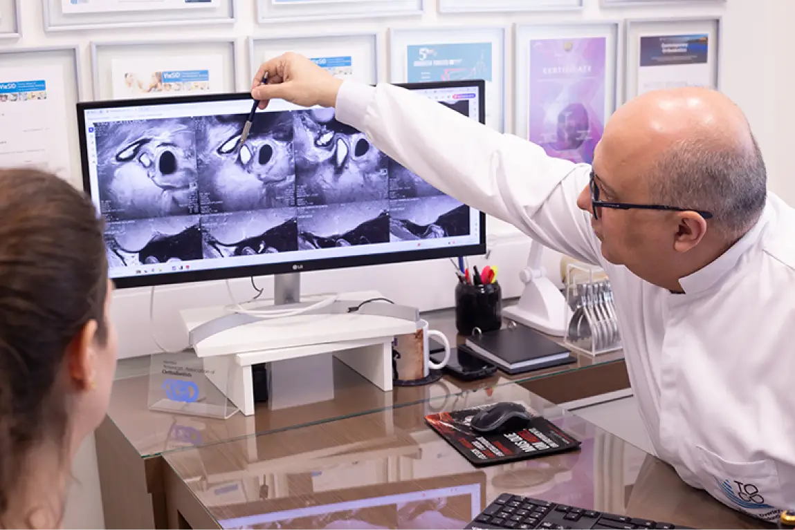
Examination with Cast Models or Digital Scanning
- Impressions of the dental arches can be made for a more detailed analysis. These models allow the dentist to precisely examine the relationship between the teeth, occlusion, and joint, enabling necessary adjustments.
- 3D scanning of the dental arches is an increasingly used technology, allowing a digital evaluation of occlusal relationships.
Use of Myorelaxant Splints
- In some cases, occlusal splints are recommended to determine the best occlusal position and relax the facial muscles, allowing a more accurate analysis of the occlusal pattern in a muscle-relaxed state.
Signs of an Inadequate Occlusal Pattern
Dental Wear
- Premature contacts or imbalances in the distribution of masticatory forces can cause irregular wear, cracks, or fractures in the teeth.
Muscle and Joint Pain
- An unbalanced occlusal pattern can overload the masticatory muscles and TMJ, leading to pain, muscle fatigue, clicking, or locking of the joint.
Headaches and Cervical Pain
- Tension from inadequate occlusion can radiate to other regions, such as the head and neck, causing tension headaches or cervical pain.
Tooth Mobility
- Improper contacts may loosen teeth, increasing the risk of mobility and, in severe cases, tooth loss.
Jaw Deviation
- In an unbalanced occlusion, the jaw may shift laterally or forward and backward during movements, causing discomfort or asymmetrical wear of the teeth.
Treatment for Occlusal Pattern Correction
Occlusal Adjustment
- Selective grinding of teeth can be performed to eliminate premature contacts and improve the distribution of masticatory forces.
Orthodontics
- In cases of malocclusion, orthodontic treatment may be recommended to reposition the teeth and improve occlusal alignment.
Occlusal Splints
- Myorelaxant or stabilizing splints can be used to relieve muscle and joint overload, protecting the teeth from excessive wear.
Prosthetic Rehabilitation
- When there is tooth loss or excessive wear, prosthetic restorations (such as crowns, veneers, or dentures) may be indicated to restore occlusal function.
Conclusion
The assessment of the occlusal pattern is a crucial step in diagnosing and treating various dental conditions, such as bruxism, temporomandibular dysfunction, and occlusal issues. Identifying occlusal misalignments early can prevent dental wear, muscle and joint pain, and improve the patient’s quality of life. A thorough examination using techniques such as palpation, articulating paper, and digital scanning is necessary to obtain an accurate diagnosis and determine the best treatment approach.