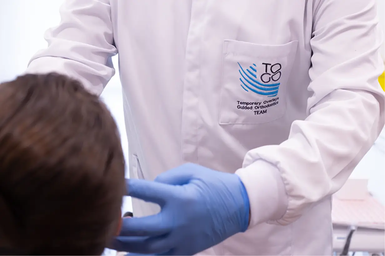
What are the main objectives of performing this exam in your clinic?
Identifying muscle pain and tension: Palpation helps detect points of tension or pain, indicating areas of muscle hyperactivity, spasms or inflammation, common in patients with bruxism or TMD.
Assessing muscle function: Checking whether the muscles are functioning properly during jaw movements, such as opening, closing and lateralization of the jaw.
Detecting asymmetries: Palpation can also identify muscle asymmetries, which may suggest imbalances in occlusion or posture.
Determining the extent of the problem: Palpation provides information about the extent of inflammation or pain, which can help guide appropriate treatment.
Muscles evaluated in dental palpation: Muscle palpation, in the dental context, usually focuses on the muscles of mastication and adjacent structures that influence the functioning of the temporomandibular joint (TMJ). The most commonly assessed muscles are:
Masseter: Located on the side of the jaw, the masseter muscle is one of the main muscles responsible for jaw closing strength. Palpation of the masseter assesses the presence of pain, hypertrophy or tension nodules.
Temporal: The temporal muscle is located on the side of the skull, with its fibers extending to the jaw. It is palpated to check for pain or tension in the temporal region, which may be related to TMJ dysfunction or dental clenching.
Medial pterygoid: The medial pterygoids are deep muscles that control opening, closing and lateral movements of the jaw. Palpation of these muscles can be more difficult, but is essential for identifying TMJ dysfunction.
Lateral pterygoid: The lateral pterygoids are deep muscles that contain two bundles: the lower one that controls anterior movements; and the upper one that controls the positioning of the articular disc. Palpation of these muscles can be more difficult, but is essential for identifying TMJ dysfunction.
Digastric: This muscle participates in retraction movements and assists in maximum jaw opening. It is also frequently palpated to identify possible radiating pain or tension associated with TMD.
Mylohyoid: The mylohyoid muscle is an important muscle in the submandibular region, forming the floor of the mouth. It extends from the mandibular region to the body of the hyoid bone, below the tongue, and is located between the digastric and geniohyoid muscles. It participates in the positioning of the hyoid bone and the jaw, being a mandibular depressor that assists in lateral-lateral movements. It is frequently palpated to identify possible radiating pain due to mandibular retraction and lateral deviations of the jaw associated with TMD.
Lateral collateral ligament:
The lateral collateral ligament is a limiter that assists in condylar centralization in the lateral-lateral direction, in relation to the articular fossa. It is also frequently palpated to identify possible radiating pain due to mandibular retraction and lateral deviations of the jaw associated with TMD.
Temporomandibular ligament: The temporomandibular ligament is an aid to the health of the temporomandibular joint, offering one of the only resistances to posterior movement of the jaw in our fragile temporomandibular joint. Palpated to identify possible radiating pain due to the retrusive positioning of the jaw associated with TMD.
Sternocleidomastoid (SCM): Although it is not a direct part of the masticatory muscles, it is frequently palpated, as tensions in these muscles may be associated with postural problems that affect the temporomandibular joint.
Trapezius: Like the sternocleidomastoid, the trapezius may present tensions associated with TMD and postural imbalances. Palpating the trapezius can help identify muscle overload, which may be related to abnormal jaw function.
How muscle palpation is performed:
Anatomical location: the dentist locates the muscle to be evaluated, using his/her anatomical knowledge to find specific points.
Controlled pressure: the pressure exerted by the fingers must be controlled and appropriate for the patient, so that sensitive areas can be detected without causing excessive discomfort. Typically, a pressure of 0.5 kgf is applied to superficial muscles and 0.25 kgf to deeper muscles.
Requesting patient feedback: during palpation, the dentist asks the patient if there is pain, discomfort or sensitivity in the palpated points. This feedback is essential for diagnosis.
Assessment of resistance and mobility: the dentist may ask the patient to perform jaw movements (such as opening and closing the mouth or moving the jaw laterally) while palpating, to observe muscle functionality.
Identifying trigger points: in some cases, the dentist can identify trigger points (areas of intense pain) that may be responsible for referred pain in other regions, such as headaches or radiating pain in the neck.
Importance of muscle palpation in dental diagnosis:
Temporomandibular Dysfunction (TMD): muscle palpation is one of the main techniques for identifying TMD. Pain and sensitivity in the masticatory muscles, especially in the masseter and temporalis, are common signs of TMD.
Bruxism: patients with bruxism often have hypertrophied and painful masticatory muscles, especially the masseter. Palpation helps to identify these muscle changes, which can be an important indicator in the diagnosis.
Orofacial pain: in cases of orofacial pain, palpation can help determine whether the origin of the pain is muscular, joint or of another nature.
Treatment planning: muscle assessment helps direct treatment, whether focused on occlusal rehabilitation, physical therapy, use of occlusal splints, muscle relaxation techniques or postural adjustments. Conclusion: Muscle palpation is an indispensable technique in dental evaluation, especially for patients with complaints of TMD, bruxism and orofacial pain. This assessment allows the professional to identify muscle changes, such as pain, tension and asymmetry, which are essential for accurate diagnosis and definition of effective treatments. The appropriate use of palpation, together with other clinical and complementary exams, offers a comprehensive view of the health of the stomatognathic system.


